68 Main Street
Irvington, NY 10533
T: 914-231-7557
F: 914-231-7558
info@squintoptometry.com Patient Registration Form
OFFICE HOURS:
The Anatomy of Your Eye
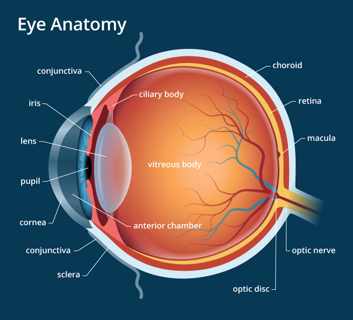
Choroid- A thin layer of tissue that is part of the middle layer of the wall of the eye, between the sclera (white outer layer of the eye) and the retina (the inner layer of nerve tissue at the back of the eye). The choroid is filled with blood vessels that bring oxygen and nutrients to the eye.
Cones- These light-sensitive nerve cells are located in the macula, which is the darkest red spot at the center of the retina. Cones are necessary for focused central vision. Cones also enable you to see colors in bright-light conditions. Routine screening photos are used to examine this area of the retina which is where macular degeneration occurs.
Conjunctiva- The conjunctiva is the clear, thin membrane that covers part of the front surface of the eye and the inner surface of the eyelids. This is a common place for eye infections to occur.
Cornea- Light first enters the eye through this transparent, dome-shaped surface that covers the front of the eye. The cornea bends, or refracts the light onto the eye’s lens. Contact lenses sit on the cornea and everyone’s cornea has a different shape. This is why contact lenses need to be fitted and it’s not one shape for everyone. It is also the part of the eye that is damaged from contact lens over wear and should be checked annually.
Iris- The iris, or the colored part of the eye surrounding the pupil, controls how much light enters the eye. The iris can make the pupil bigger or smaller by opening or closing. This is what is affected by dilating drops during a dilated eye exam.
Lens- Behind the pupil and the iris is a transparent structure that looks similar in shape to the lens of a magnifying glass. Unlike glass lenses, though, this part of the eye can change shape. This enables it to bend the rays of light even more, so they land in the right place on the retina, at the back of the eye. The lens gets more yellow as it ages, and sometimes gets opaque spots in it, which is a cataract. This is the part of the eye that is removed and replaced during cataract surgery.
Macula- The macula is the small area at the center of the retina responsible for what we see straight in front of us. It is the most pigmented part of the retina and the area of the eye affected by age related macular degeneration.
Optic nerve- The cells of the retina turn light into electrical impulses. These electrical signals are collected by the optic nerve, a bundle of about 1 million nerve fibers, and transmitted to the brain. The brain puts all this information together to produce the image that you see. The optic nerve can change from diseases like glaucoma and multiple sclerosis. Screening photos help determine if there is a change in the optic nerve.
Pupil- This is the round hole at the front of the eye that appears black. It is located behind the middle of the cornea and is surrounded by the iris. It is a large black hole that looks smaller in bright light and larger in dark light when the eye is dilated.
Retina- At the back of the eye is the retina, or a thin layer of light-sensitive nerve cells. The retina contains different types of photoreceptors, called rods and cones, which respond to light that lands on them. Different diseases like diabetes and hypertension affect the health of the retina. This is why annual eye exams, including dilation, are important.
Rods-These light-sensitive nerve cells surround the macula and extend to the edge of the retina. The rods provide you with your side, or peripheral vision. They also help you see at night and in dim light. A visual field test is used to access your peripheral vision, which can decrease with glaucoma.
Sclera- The sclera is the part of the eye commonly known as the “white.” It forms the supporting wall of the eyeball, and is continuous with the clear cornea. The sclera is covered by the conjunctiva.
Vitreous body/Gel- The eye is filled with a gel that helps it keep its round shape. Light entering the eye first passes through the cornea then the lens and then the vitreous body before reaching the retina. Floaters are bits of material that float in the vitreous, casting a shadow on the retina and causing you to see a spot in your vision.